Tarepia celular e incontinência urinária
A incontinência urinária (IU) afeta mais de 200 milhões de pessoas em todo o mundo (Norton, Brubaker, 2006). Incontinência urinária de esforço (IUE) é o tipo mais comum em mulheres, sendo responsável por aproximadamente metade dos casos de IU, com a maioria dos estudos reportando prevalência de 10 a 39% (Abrams et al., 2013).
O tratamento da IUE pode ser dividido em clínico e cirúrgico, porém nenhuma das terapias convencionais se concentra na parada de progressão da doença ou na reversão do dano existente. Assim, a medicina regenerativa surgiu como um emocionante meio de preencher esse vazio terapêutico através da restauração e manutenção da função normal, com efeitos diretos em tecidos lesados ou disfuncionais (Furuta et al. 2007; Wang et al., 2011; Goldman et al., 2012; Vaegler et al., 2012).
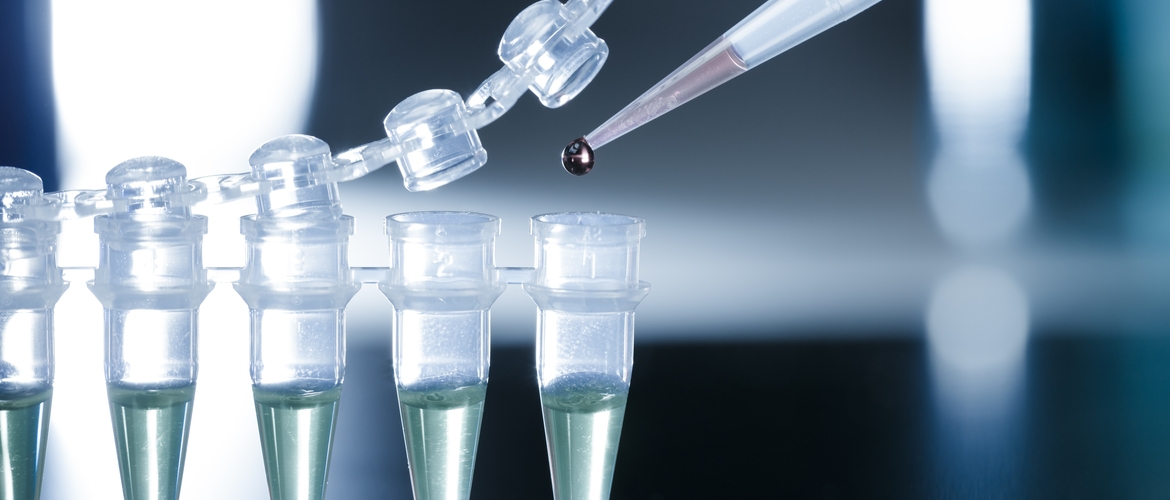
As células-tronco são classicamente definidas por sua capacidade de auto-renovação, proliferação e diferenciação em um ou mais tipos de células a longo prazo (Ho, Bhatia, 2012). Assim, tipos de células-tronco diferentes possuem diferentes habilidades.
Células-tronco toti ou pluripotentes, tipicamente embrionárias ou fetais na origem podem se diferenciar em qualquer tipo de célula ou camada celular germinativa. As multipotentes, incluindo células-tronco mesenquimais (a partir de sangue, medula óssea, placenta, tecido adiposo ou muscular) são capazes de se diferenciar em vários, mas não todos, tipos celulares. As células-tronco unipotentes (de pele por exemplo) são as mais limitadas, sendo capazes de regeneração em apenas um tipo celular (Fong et al., 2010; Ho, Bhatia, 2012; Singh, 2012).
Atualmente, quatro grandes categorias descrevem a diversidade de células-tronco investigadas em medicina regenerativa: células-tronco embrionárias (ESC), células derivadas da placenta ou do líquido amniótico (AFPs), células pluripotentes induzida (IPSC), e células-tronco adultas (ASC) (Martin, 1981; Thomson et al., 1998; Goldman et al., 2012; Vaegler et al., 2012; Kim et al., 2013).
O conceito inicial da terapia celular para IUE foi baseado na hipótese de que a injeção de mioblastos esqueléticos na uretra lesionada pode restaurar o músculo do esfíncter (Chancellor et al., 2000). Desde então evoluiu com a substituição dos mioblastos por outros tipos de células, como as células-tronco derivadas de músculo (MDSC), de adipócitos (ADSC), de medula óssea (BMSC) e AFPs.
Assim, diversos estudos pré-clínicos demonstraram melhora funcional com aumento da pressão de perda (LPP) na cistometria, melhora da amplitude de contração rápida uretral e melhora histológica uretral (Lee et al., 2003; Cannon et al., 2003; Chermansky et al., 2004; Kwon et al., 2006; Furuta et al., 2008; Xu et al., 2010; Lin et al., 2010; Wu et al., 2011; Zhao et al., 2011; Watanabe et al., 2011), servindo como base para o início das pesquisas em humanos.
A terapia celular em mulheres com IUE foi aplicada pela primeira vez por Mitterberger, Marksteiner et al. (2007a). Para avaliar sua eficácia, eles usaram o questionário I-QOL, teste urodinâmico e ultrassom para visualizar o rabdoesfincter e avaliar sua contratilidade. Ultrassom mostrou que o rabdoesfincter tornou-se mais espesso e sua contratilidade melhorou um ano após injeção de mioblastos e fibroblastos. A Pressão máxima de fechamento uretral na avaliação urodinâmica (PMFU) melhorou 40,6% em 119 mulheres.
Como foi o caso de outros estudos clínicos (Mitterberger, Pinggera et al., 2008b; Sebe et al., 2011), entre os critérios de exclusão foram destacados hipermobilidade uretral e prolapso do colo vesical, pois podem causar incontinência em pessoas cuja função esfincteriana está intacta.
Uma série de estudos que utilizaram diferentes questionários (I-QOL, ICIQ-QOL, PGI-I, etc.) demonstraram boa eficácia das terapias celulares em mulheres (Blaganje & Lukanović, 2012; Carr et al., 2013; Gras et al., 2014); no entanto, os achados desses estudos devem ser interpretados com cautela, pois não foram avaliados usando testes urodinâmicos.
Ao mesmo tempo, Kuismanen et al. (2014) não encontraram aumento na PMFU após injeções de ADSCs combinadas com gel de colágeno, o que pode ser devido a um curto período de seguimento (6 meses) ou a um pequeno número de pacientes (n = 5).
Dois estudos de viabilidade avaliaram a eficácia potencial de diferentes doses de células, bem como diferentes quantidades de doses, e sugeriram que as injeções de maior dose estão associadas a uma melhora maior da IUE (Carr et al., 2013; Peters et al., 2014). No entanto, ambos utilizaram apenas métodos de avaliação subjetiva e nenhum mencionou efeitos secundários estatisticamente relevantes, indicando segurança relativa do tratamento.
Assim, a terapia com células-tronco representa uma modalidade promissora de tratamento para IUE, tendo sua indicação em pacientes com defeito esfincteriano intrínseco, podendo ser uma boa opção futura em pacientes com uretra fixa. Entretanto, antes de se tornar uma prática clínica mais estudos são necessários para elucidar sua eficácia a longo prazo e segurança, assim como a dose de células, número de doses, via de administração e o tipo de célula-tronco ideal.
Referências:
Abrams P, Cardozo L, Khoury S, Wein A. Incontinence (5th Edition) 2013, Health Publication Ltd, Paris, France.
Blaganje, M, & Lukanović, A. Intrasphincteric autologous myoblast injections with electrical stimulation for stress urinary incontinence. International Journal of Gynaecology and Obstetrics: The Official Organ of the International Federation of Gynaecology and Obstetrics. 2012; 117(2), 164–167.
Cannon TW, Lee JY, Somogyi G, et al. Improved sphincter contractility after allogenic muscle-derived progenitor cell injection into the denervated rat urethra. Urology. 2003; 62:958-63.
Carr, LK, Robert, M, Kultgen, PL, et al. Autologous muscle derived cell therapy for stress urinary incontinence: A prospective, dose ranging study. Journal of Urology. 2013; 189(2), 595–601.
Chancellor MB, Yokoyama T, Tirney S, et al. Preliminary results of myoblast injection into the urethra and bladder wall: a possible method for the treatment of stress urinary incontinence and impaired detrusor contractility. Neurourol Urodyn. 2000;19(3):279-87.
Chermansky CJ, Tarin T, Kwon DD, et al. Intraurethral muscle-derived cell injections increase leak point pressure in a rat model of intrinsic sphincter deficiency. Urology. 2004; 63:780-5.
Fong CY, Gauthaman K, Bongso A. Teratomas from pluripotent stem cells: a clinical hurdle. J Cell Biochem. 2010; 111:769-81.
Furuta A, Jankowski R, Honda M, et al. State of the art of where we are at using stem cells for stress urinary incontinence. Neurourol Urodyn. 2007; 26:966-71.
Goldman H, Sievert K, Damaser M. Will we ever use stem cells for the treatment of SUI?-ICI-RS 2011. Neurourol Urodyn. 2012; 31:386-9.
Gras, S., Klarskov, N., & Lose, G. Intraurethral injection of autolo- gous minced skeletal muscle: A simple surgical treatment for stress urinary incontinence. Journal of Urology. 2014; 192(3), 850–855.
Ho CP, Bhatia NN. Development of stem cell therapy for stress urinary incontinence. Curr Opin Obstet Gynecol. 2012; 24:311-7
Kim J, Lee S, Song Y, Lee H. Stem cell therapy in bladder dysfunction: where are we? And where do we have to go? Biomed Res Int. 2013:930713.
Kuismanen, K, Sartoneva, R, Haimi, S, et al. Autologous adipose stem cells in treatment of female stress urinary incontinence: Results of a pilot study. Stem Cells Translational Medicine. 2014; 3(8), 936–941.
Kwon D, Kim Y, Pruchnic R, et al. Periurethral cellular injection: comparison of muscle-derived progenitor cells and fibroblasts with regard to efficacy and tissue contractility in an animal model of stress urinary incontinence. Urology. 2006; 68:449-54.
Lee JY, Cannon TW, Pruchnic R, et al. The effects of periurethral muscle-derived stem cell injection on leak point pressure in a rat model of stress urinary incontinence. Int Urogynecol J Pelvic Floor Dysfunct. 2003;14:31-7.
Lin G, Wang G, Banie L, et al. Treatment of stress urinary incontinence with adipose tissue-derived stem cells. Cytotherapy. 2010; 12:88-95.
Martin G. Isolation of a pluripotent cell line from early mouse embryos cultured in medium conditioned by teratocarcinoma stem cells. Proc Natl Acad Sci USA. 1981; 78:7634-8.
Mitterberger, M, Marksteiner, R, Margreiter, E, et al. Autologous myoblasts and fibroblasts for female stress incontinence: A 1‐year follow‐up in 123 patients. BJU International. 2007; 100(5), 1081–1085.
Mitterberger, M, Pinggera, GM, Marksteiner, R, et al. Adult stem cell therapy of female stress urinary incontinence. European Urology. 2008; 53(1), 169–175.
Norton P, Brubaker L. Urinary incontinence in women. Lancet. 2006; 367:57-67.
Peters, KM, Dmochowski, RR, Carr, LK, et al. Autologous muscle derived cells for treatment of stress urinary incontinence in women. Journal of Urology. 2014; 192(2), 469–476.
Sebe, P, Doucet, C, Cornu, JN, et al. Intrasphincteric injections of autologous muscular cells in women with refractory stress urinary incontinence: A prospective study. International Urogynecology Journal. 2011; 22(2), 183–189.
Singh SR (Ed). Somatic stem cells: methods and protocols. New York: Humana Press, Springer; 2012.
Thomson J, Itskovitz-Eldor J, Shapiro S, et al. Embryonic stem cell lines derived from human blastocysts. Science. 1998; 282:1145-7.
Vaegler M, Lenis A, Daum L, et al. Stem cell therapy for voiding and erectile dysfunction. Nat Rev Urol. 2012; 9:435-47.
Wang H, Chuang Y, Chancellor M. Development of cellular therapy for the treatment of stress urinary incontinence. Int Urogynecol J. 2011; 22:1075-83.
Watanabe T, Maruyama S, Yamamoto T, et al. Increased urethral resistance by periurethral injection of low serum cultured adipose-derived mesenchymal stromal cells in rats. Int J Urol. 2011;18(9):659-66.
Wu S, Wang Z, Bharadwaj S, et al. Implantation of autologous urine derived stem cells expressing vascular endothelial growth factor for potential use in genitourinary reconstruction. J Urol. 2011; 186:640-7.
Zhao W, Zhang C, Jin C, et al. Periurethral injection of autologous adipose-derived stem cells with controlled-release nerve growth factor for the treatment of stress urinary incontinence in a rat model. Eur Urol. 2011 Jan;59(1):155-63.
Autores:
Andreisa Paiva M Bilhar1 e Rodrigo de Aquino Castro2
- 1- Doutora em Ginecologia pela Universidade Federal de São Paulo. Membro da Comissão Nacional Especializada Uroginecologia e Cirurgia Vaginal- FEBRASGO
- 2- Professor Associado Livre Docente do Departamento de Ginecologia da Unifesp – EPM. Presidente da Comissão Nacional Especializada em Uroginecologia e Cirurgia Vaginal- FEBRASGO


